
Everything You Need to Know About Melanoma
Melanoma is a type of cancer of the skin. It starts in cells called melanocytes in the skin. Melanocytes are cells that make melanin, which is what gives your skin its colour.
Melanomas only make up about 1 percent of skin cancers. Melanoma is also called malignant melanoma or cutaneous melanoma.
Most people do well with treatment for melanoma when it is found early on. But if it is not caught early, it is easy for the disease to spread to other parts of the body.
What are the symptoms?
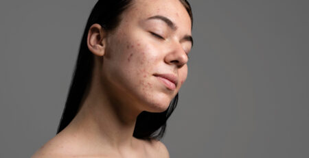
Melanoma’s first signs and symptoms are:
- changes made to a mole
- the growth of a new, strange thing on your skin
If melanin is still being made by melanoma cells, the tumours are usually brown or black. Some melanomas don’t make melanin, so these tumours can be tan, pink, or white.
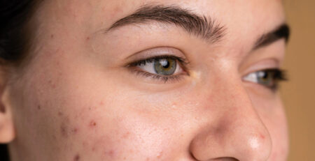
Melanoma could be present in a mole if:
- irregular shape
- irregular border
- multicolored or uneven coloring
- larger than a quarter of an inch
- changes in size, shape, or color
- itchiness or bleeding
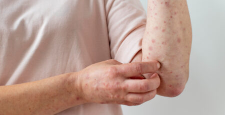
Melanoma can begin anywhere on your skin. But the most likely places are:
- chest and back for men
- legs for women
- neck
- face
This could be because these parts of the body get more sunlight than other parts. Melanoma can grow in places that don’t get much sun, like the palms of your hands, the soles of your feet, and the beds of your fingernails.
Types of melanoma
The most common kind of melanoma is one that spreads on the skin’s surface. It usually spreads across the skin’s surface, has uneven edges, and can be brown, black, pink, or red.
Nodular melanoma is another type that grows deeper into the skin and can look like a raised bump or growth.
Lentigo maligna melanoma usually shows up on the face and other parts of the body that get more sun, especially in older people. It looks like a big, bumpy, dark patch on the skin’s surface.
Melanoma that has spread, or metastasized, to other parts of the body, like the lymph nodes, organs, or bones, is called metastatic melanoma.
There are also other rare types of melanoma. Most of the time, it affects the skin, but some types can also affect internal tissues and the eyes.
Mucosal melanoma can form in the mucous membranes that line:
- digestive tract
- mouth
- nose
- urinary tract
- vagina
Melanoma of the eye, also called ocular melanoma, can happen under the white part of the eye.
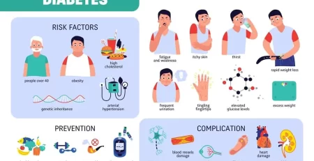
Risk factors
Ultraviolet light
It’s not completely clear what causes melanoma, but exposure to the sun and other sources of ultraviolet light, like tanning beds, is a very important risk factor.
Race
The American Cancer Society says that the risk of getting melanoma over the course of a lifetime is about:
- 2.6% for white people
- 0.1% for Black people
- 0.6% for Hispanic people
It is said that white people get melanoma 20 times more often than Black people. Keep in mind that this data could be the result of differences in healthcare and other factors.
Along with having lighter skin, having a lot of moles may also put you at risk.
Genetics/family history
If a parent or sibling has had melanoma before, you may be more likely to get it yourself.
Age
Melanoma is more likely to happen as you get older. Even though it is one of the most common cancers in young adults, the average age when it is found is 65.
What are the stages of melanoma?
Staging cancer shows how far the cancer has spread from where it started. Cancer can spread to other parts of the body through the tissues, the lymph system, and the bloodstream.
Melanoma has these stages:
Stage 0
You have weird melanocytes, but they are only on the top layer of skin (epidermis). Melanoma in situ is another name for this.
Stage 1
- 1A: You have a tumour that is cancerous, but it is less than 1 mm thick. It doesn’t have any sores.
- 1B: The tumour is less than 1 mm thick, but it has sores on it. Or, it’s between 1 and 2 mm thick and there are no sores.
Stage 2
- 2A: The tumor is between 1–2 mm thick with ulceration. Or, it’s between 2–4 mm thick without ulceration.
- 2B: The tumour is between 2 and 4 mm in size and has holes in it. Or it is thicker than 4 mm and doesn’t have any ulcers.
- 2C: The tumor is over 4 mm thick and is ulcerated.
Stage 3
You have a tumor of any size that may or may not be ulcerated. At least one of these is also true:
- At least one lymph node has been found to have cancer.
- The lymph nodes are connected.
- Between the tumour and the closest lymph nodes, cancer was found in a lymph vessel.
- More than 2 cm away from the main tumour, cancer cells have been found.
- Within 2 cm of the main tumour, other small ones have been found on or under your skin.
Stage 4
The cancer has spread to places far away. This can include organs, soft tissue, and bone.
What’s the treatment?
The treatment for melanoma depends on how far along it is.
Stage 0
Melanoma in stage 0 is only in the top layer of skin. During a biopsy, it is possible to get rid of all of the suspicious tissue. If it doesn’t, your doctor can cut it out along with a small border of healthy skin.
You might not need any more care.
Stage 1 and 2
During a biopsy, very thin melanomas can be taken out completely. If not, they can be taken out with surgery later. This means taking out the cancer along with some healthy skin around it and a layer of tissue under the skin.
Melanoma in its early stages doesn’t always need more treatment.
Stage 3 and 4
Melanoma in stage 3 has spread outside of the main tumour or to lymph nodes nearby. Wide-excision surgery is used to get rid of the tumour and the lymph nodes that are affected by it.
In stage 4, the cancer has spread to places far away. Surgery can remove the skin tumours and some of the swollen lymph nodes. Tumors on organs inside your body can also be taken out by surgery. But your surgery options depend on how many tumours you have, how big they are, and where they are.
Most of the time, extra treatments are needed for stages 3 and 4, which may include:
- Drugs for immunotherapy. Interferon, interleukin-2, and checkpoint inhibitors like ipilimumab (Yervoy), nivolumab (Opdivo), and pembrolizumab are examples of these (Keytruda).
- Targeted treatment for cancers that are caused by changes in the BRAF gene. Some of these are cobimetinib (Cotellic), dabrafenib (Tafinlar), trametinib (Mekinist), and vemurafenib (Zelboraf).
- Melanoma caused by changes in the C-KIT gene can be treated with targeted therapy. Imatinib (Gleevec) and nilotinib are two examples (Tasigna).
- Vaccines. Bacille Calmette-Guerin (BCG) and T-VEC are two of these (Imlygic).
- Therapy with radiation. This can be used to shrink tumours and kill cancer cells that surgery might have missed. Cancer that has spread to other parts of the body can also be helped by radiation.
- Isolated perfusion of a limb. This is done by putting a heated solution of chemotherapy into only the affected arm or leg.
- Systemic treatment. This could include the drugs dacarbazine (DTIC) and temozolomide (Temodar), which can be used to kill cancer cells all over your body.
Immunotherapy and targeted therapies can’t cure melanoma, but they can make people live longer. Melanoma chemotherapy can shrink tumours, but they can come back after a few months.
Each type of therapy has its own set of side effects, some of which can be serious. It’s important to talk to your doctor about these things so you can make a smart choice.
Clinical trials can help you get new treatments that haven’t been approved for use by everyone else yet. Talk to your doctor if you want to take part in a clinical trial.
What causes melanoma?
Usually, healthy new skin cells push older skin cells to the surface, where they die.
Damage to the DNA in the melanocytes can make new skin cells grow too fast. As the skin cells pile up, they make a lump called a tumour.
It’s not clear why skin cells’ DNA gets damaged. It could be a mix of things like genes and the environment.
Exposure to ultraviolet (UV) radiation may be the main cause. UV rays can come from the sun, tanning beds, and tanning lamps, among other places.
How is it diagnosed?
Physical examination
First, you’ll have to look closely at your skin. As adults, most of us have between 10 and 40 moles.
A normal mole is usually the same colour all over and has a clear edge. They are usually less than a quarter of an inch in diameter and can be round or oval.
A good skin exam will include looking in places that are less obvious, such as:
- between the buttocks
- genitals
- palms and under your fingernails
- scalp
- soles of your feet, between your toes, and under your toenails
Blood chemistry studies
Your doctor can look for lactate dehydrogenase in your blood (LDH). When you have melanoma, you may have more of this enzyme than usual.
LDH levels may not be checked to find diseases in their early stages.
Skin biopsy
A skin biopsy is the only way to be sure melanoma is present. During a biopsy, a small piece of skin is taken for testing. If at all possible, the whole area under suspicion should be taken away. The tissue is then sent to a lab where it is looked at with a microscope.
Your doctor will get the pathology report and explain what it means.
If melanoma is found, it is important to find out what stage it is in. This will tell you about your overall outlook and help plan your treatment.
The first step in staging is to measure the size of the tumour. A microscope can be used to measure the size of the melanoma.
Lymph node biopsy
If you have been diagnosed, your doctor may need to check to see if the cancer cells have spread. If you have melanoma in situ, they won’t do this. The first thing to do is a sentinel node biopsy.
For the surgery, the area where the tumour was will be injected with a dye. This dye will naturally go to the lymph nodes that are close by. The lymph nodes will be taken out by the surgeon so they can be checked for cancer.
If there is no cancer in the sentinel nodes, it is likely that the cancer hasn’t spread beyond the area that was tested at first. If there is cancer, the next set of lymph nodes may be checked.
Imaging tests
Imaging tests are done to find out if skin cancer has spread to other parts of the body.
- A CT scan. Before the scan, a dye will be put into one of your veins. X-rays will be taken from different points of view. The dye will help organs and tissues stand out.
- MRI. A substance called gadolinium is put into a vein and used for this test. The scanner takes pictures with a magnet and radio waves, and the gadolinium makes cancer cells stand out.
- PET scan. A small amount of radioactive glucose must be put into a vein for this test. The scanner will then move around you. Because they use more glucose, cancer cells stand out on the screen.
Melanoma survival rates
It makes sense to want to look into survival rates, but it’s important to remember that they’re just averages. Your circumstances are unique to you, so speak to your doctor about your own prognosis.
Based on data from 2010 to 2016, the 5-year relative survival rates for melanoma of the skin in the United States is 92.7 percent overall, and:
- 99% for localized melanoma
- 66.3% for regional spread
- 27.3% for distant metastasis
Melanoma is found at the local stage about 83 percent of the time.
Prevention tips
Even though you can’t completely avoid risk, here are a few things you can do to help stop melanoma and other skin cancers:
- If you can, stay out of the sun during the middle of the day. Remember that even on cloudy days and in the winter, the sun can still hurt your skin.
- Use sunscreen. Use a sunscreen with at least 30 SPF that covers a wide range of wavelengths. Apply more sunscreen every two hours or more often if you sweat a lot or go in the water. Do this no matter what time of year it is.
- Stealth. When you’re outside, make sure your arms and legs are covered. Wear a hat with a wide brim to keep your head, ears, and face safe.
- Put on sunglasses that block UVA and UVB rays.
- Don’t use tanning lamps or beds.
What’s the outlook?
Survival rates can only give you a rough idea of your own chances. Your doctor can give you a more personal evaluation.
Some things that can change how you feel are:
- Age. People who are older tend to live less long.
- Health in general. Your treatment may not work as well if your immune system is weak or if you have other health problems.
From the relative survival rates listed above, you can see that many people with melanoma do survive. Melanoma in its later stages is harder to treat, but it is still possible to live for many years after it is found.
Every year, 22 out of every 100,000 people in the United States are told they have melanoma. The better your chances are, the sooner it’s found and treated.
You may have a better chance of getting a diagnosis early if:
- Check your body often for any changes. Note any changes in the size, shape, or colour of moles, freckles, and birthmarks that you already have. Check the bottoms of your feet, the spaces between your toes, and the nail beds. Use a mirror to check places like your genitalia and the space between your buttocks that are hard to see. Take pictures so you can see changes more easily. And tell your doctor right away about anything that seems odd.
- See your primary care doctor once a year for a full checkup. If your doctor doesn’t check your skin, request it. Or, ask for a dermatologist’s name.


Leave a Reply